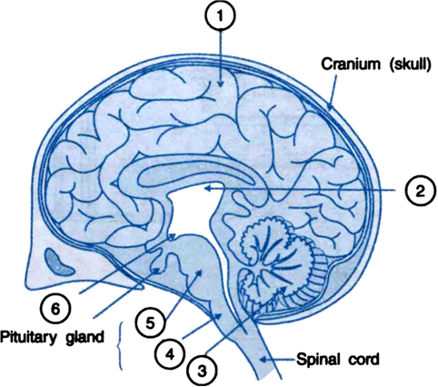
40 the brain with labels
Brain: Ultimate Guide to the Brain for AP® Psychology Luckily, you stumbled across this ultimate guide to the brain for AP® Psychology that we have prepared for you. In this AP® Psychology crash course review, we will provide a summary of the anatomy and function of the major areas of the brain. The brain is divided into three main parts: the forebrain, the midbrain, and the hindbrain. Anatomy of the Brain | Simply Psychology It consists of grey matter (the cerebral cortex) and white matter at the centre. The cerebrum is divided into two hemispheres, the left and right, and contains the lobes of the brain (frontal, temporal, parietal, and occipital lobes). The cerebrum produces higher functioning roles such as thinking, learning, memory, language, emotion, movement ...
Can a Label Shortage Bring Global Supply Chains to a Halt ... A significant shortage in these types of labels threatens to bring a supply chain already strained by inflation, war and the COVID-19 pandemic to a standstill. While China is by far the largest producer of paper and cardboard, Finland ranks seventh, with two Finnish companies, UPM and Stora Enso, among the top seven companies producing paper ...

The brain with labels
Labeled imaging anatomy cases | Radiology Reference ... URL of Article. This article lists a series of labeled imaging anatomy cases by body region and modality. On this page: Article: Brain. Head and neck. Spine. Chest. Abdomen and pelvis. Cross-sectional anatomy of the brain - e-Anatomy - IMAIOS Anatomy of the brain: how to view anatomical labels. This module is a comprehensive and affordable learning tool for medical students and residents and especially for neuroradiologists and radiation oncologists. It provides access to an atlas and to images in axial planes, allowing the user to learn and review neuroanatomy interactively. ... Brain: Atlas of human anatomy with MRI - e-Anatomy Anatomy of the brain (MRI) - cross-sectional atlas of human anatomy. The module on the anatomy of the brain based on MRI with axial slices was redesigned, having received multiple requests from users for coronal and sagittal slices. The elaboration of this new module, its labeling of more than 524 structures on 379 MRI images in three different ...
The brain with labels. Children's products labeled water- or stain-resistant may ... April 20, 2022 — Tiny capsule that delivers CRISPR gene therapy to the brain could be used to target glioblastoma tumours. Meningitis vaccine may be a new weapon against 'super-gonorrhoea' PICASSO allows ultra-multiplexed fluorescence imaging of ... For each staining and imaging round, a brain slice was stained with 15 different preformed antibody complexes that were labeled with 15 spectrally overlapping fluorophores and then it was imaged ... Can the Brain Predict Future Risk for Psychological ... Scientists have turned to the brain for answers on the biological bases of these varied trauma responses and to discover brain-based profiles that are agnostic to diagnostic labels. Parts of the Human Brain | Anatomy & Function - Video ... The diagram shows the brain anatomy with labeled parts of the human brain. Lesson Summary. The brain is the control center of the body. It is in charge of everything from thought, memory, and ...
Innovative Brain-Wide Mapping Reveals a Single Memory Is ... Innovative brain-wide mapping study shows that "engrams," the ensembles of neurons encoding a memory, are widely distributed, including among regions not previously realized. A new study from MIT 's Picower Institute for Learning and Memory provides the most extensive and rigorous evidence yet that the mammalian brain retains a single ... Free Nervous System Worksheets and Printables Brain Hemisphere Chart - Make a paper hat to wear that shows the main parts of the brain! Your kids will love seeing where the brain lobes are located under their skulls. Label the Brain Anatomy Diagram - This brain anatomy labeling worksheets is a great addition to the study of human anatomy. Researchers uncover how Listeria infects the brain | Food ... The central nervous system is separated from the bloodstream by what is known as the blood-brain barrier. But some pathogens manage to cross it and are able to infect the central nervous system. Brain: Anatomy, Pictures, Functions, and Conditions The Brain Stem. PIXOLOGICSTUDIO/SCIENCE PHOTO LIBRARY / Getty Images. The brainstem is an area located at the base of the brain that contains structures vital for involuntary functions such as the heartbeat and breathing. The brain stem is comprised of the midbrain, pons, and medulla. 3.
Parts of the Brain: Structures, Anatomy and Functions The brain is a 3-pound organ that contains more than 100 billion neurons and many specialized areas. There are 3 main parts of the brain include the cerebrum, cerebellum, and brain stem.The Cerebrum can also be divided into 4 lobes: frontal lobes, parietal lobes, temporal lobes, and occipital lobes.The brain stem consists of three major parts: Midbrain, Pons, and Medulla oblongata. Brain: Function and Anatomy, Conditions, and Health Tips The brain is an organ that's made up of a large mass of nerve tissue that's protected within the skull. It plays a role in just about every major body system. Some of its main functions ... Positions and Functions of the Four Brain Lobes | MD ... The occipital lobe, the smallest of the four lobes of the brain, is located near the posterior region of the cerebral cortex, near the back of the skull. The occipital lobe is the primary visual processing center of the brain. Here are some other functions of the occipital lobe: Visual-spatial processing. Movement and color recognition. Third harmonic generation imaging for fast, label-free ... (D) Infrared photons (white arrow) are focused deep in the brain tissue, converted to THG (green) and SHG (red) photons, scattered back (green/red arrows) and epi-detected. The nonlinear optical processes result in label-free contrast images with sub-cellular resolution and intrinsic depth sectioning.
Parts of the brain: Learn with diagrams and quizzes | Kenhub Labeled brain diagram. First up, have a look at the labeled brain structures on the image below. Try to memorize the name and location of each structure, then proceed to test yourself with the blank brain diagram provided below. Labeled diagram showing the main parts of the brain.
Template anatomical atlases and parcellation schemes ... Tags: template atlas Template anatomical atlases and parcellation schemes. An atlas is a volumetric or surface based description of the geometry of the brain, where each anatomical coordinate is labeled according to some scheme, e.g., as Brodmann area.A recent review of brain templates and atlases is presented in Brain templates and atlases (2012) in NeuroImage.
How Well Do You Know About Parts Of The Brain? - ProProfs Thalamus. D. Cerebrum. 6. The ________ lobes are in the rear of the brain and contain the visual cortex, which processes sight. 7. The lobe that is located near the ear and is in charge of hearing is the ____________ lobe. A. Occipital.
Sagittal Brain Mri Labeled - go anatomy s app tries to ... Sagittal Brain Mri Labeled. Here are a number of highest rated Sagittal Brain Mri Labeled pictures upon internet. We identified it from obedient source. Its submitted by giving out in the best field. We agree to this nice of Sagittal Brain Mri Labeled graphic could possibly be the most trending topic as soon as we ration it in google lead or ...
Ventricles of the Brain Function, Anatomy & Diagram ... A schematic diagram of the brain, with the lateral ventricles labeled. ... The brain is a very fragile organ within the human body that relies on a lot of energy in the form of glucose. For that ...
News Bureau | ILLINOIS The Illinois team wanted to use the power of MRI to directly image epigenetic changes in live subjects. For the new approach, the team relied on a key insight: Li realized that an essential amino acid, methionine, could carry an atomic marker known as carbon-13 into the brain, where it could donate the carbon-13-labeled methyl group needed for ...
Brain Ventricles: Anatomy, Function, and Conditions Function. Aside from cerebrospinal fluid, your brain ventricles are hollow. Their sole function is to produce and secrete cerebrospinal fluid to protect and maintain your central nervous system. CSF is constantly bathing the brain and spinal column, clearing out toxins and waste products released by nerve cells.
Mapping the Brain to Understand the Mind - Scientific American He was a codeveloper of Brainbow, for example—a genetic technique that can label individual neurons in hundreds of different hues, producing spectacular images of the brain. More recently, he ...
Lobes of the brain: Structure and function | Kenhub The frontal lobe is the largest lobe of the brain comprising almost one-third of the hemispheric surface. It lies largely in the anterior cranial fossa of the skull, leaning on the orbital plate of the frontal bone.. The frontal lobe forms the most anterior portion of the cerebral hemisphere and is separated from the parietal lobe posteriorly by the central sulcus, and from the temporal lobe ...
Parts of the Brain Activity for Kids, Brain Diagram, and ... This brain coloring sheet was my kids' favorite! Brain worksheet for grade 2. Parts of the Brain Worksheet - Label the human brain by writing the number on the brain template; Label the Brain Parts Worksheet - Use the brain vocabulary from the word bank to label the brain areas; Brain Activity for kids
Your guide to understanding Nutrition Facts labels | The Star The list gives you information on 13 core nutrients: fat, saturated fat, trans-fat, cholesterol, carbohydrate, sodium, fibre, sugars, protein, vitamin A, vitamin C, calcium and iron. If these ...
Brain: Atlas of human anatomy with MRI - e-Anatomy Anatomy of the brain (MRI) - cross-sectional atlas of human anatomy. The module on the anatomy of the brain based on MRI with axial slices was redesigned, having received multiple requests from users for coronal and sagittal slices. The elaboration of this new module, its labeling of more than 524 structures on 379 MRI images in three different ...
Cross-sectional anatomy of the brain - e-Anatomy - IMAIOS Anatomy of the brain: how to view anatomical labels. This module is a comprehensive and affordable learning tool for medical students and residents and especially for neuroradiologists and radiation oncologists. It provides access to an atlas and to images in axial planes, allowing the user to learn and review neuroanatomy interactively. ...
Labeled imaging anatomy cases | Radiology Reference ... URL of Article. This article lists a series of labeled imaging anatomy cases by body region and modality. On this page: Article: Brain. Head and neck. Spine. Chest. Abdomen and pelvis.




Post a Comment for "40 the brain with labels"